Bimonthly internal assessment
Case 1:
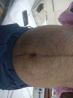
Q1 What is the Reason for this patients ascites ?
The most common cause of Ascites is
Cirrhosis of liver
risk factors in this patient :
1. Chronic alcoholism since 40 years
2. Truncal obesity leading to metabolic syndrome causing NAFLD leading to cirrhosis
https://www.ncbi.nlm.nih.gov/pmc/articles/PMC6092576/
Altered echo texture of liver due to Cirrhosis causes portal hypertension leading to increased hydrostatic pressure causing fluid accumulation hence Ascites
Q2 Bilateral pedal oedema which is of pitting type is due to decrease in the albumin level trends due to course of the disease and long standing cirrhosis causing decrease in the production of proteins causing decrease in the oncotic pressure leading to accumulation of fluid.
as per the given clinical data due to chronic liver disease there was increasing trend of INR which was as high as 4.7 causing bleeding manifestations ( bleeding gums, hematoma formation )
ulcerations are due his limited self practising manoeuvres done in inappropriate conditions such as
improper dressing of the wound, not maintaining aseptic conditions , indescriminate use of steroids (self medication) to quote from history (Patient attenders were giving history of multiple self medications whenever patient developed fever or shortness of breath they used to take decadron injections, larigo tablets,monocef and pantop over 4 months intermittently ) causing ? immune suppression leading to secondary infections hence cellulitis and non healing of wound.
Q3 Reason for Asterexis and constructional apraxia
Asterixis is a clinical sign that describes the inability to maintain sustained posture with subsequent brief, shock-like, involuntary movements.
this clinical sign is not pathognomonic for any condition. However, it may indicate a serious underlying disease process.
https://www.ncbi.nlm.nih.gov/books/NBK535445/
West haven criteria is being used for HE
In hepatic encephalopathy (due to cirrhosis of liver ) damage occurs to brain cells due to the impaired metabolism of ammonia is predominantly related to the development of asterixis in hepatic encephalopathy,
pathophysiology of HE
1 role of neurotoxins,
2.impaired neurotransmission due to metabolic changes in liver failure, changes in brain energy metabolism, systemic inflammatory response and alterations of the blood brain barrier which produces a wide spectrum of nonspecific neurological and psychiatric manifestations.
https://www.ncbi.nlm.nih.gov/pmc/articles/PMC5421503
lactulose and rifaximin
underlying mechanism of Giving lactulose
The human small intestinal mucosa does not have the enzymes to split lactulose, and hence lactulose reaches the large bowel unchanged.
basically three mechanisms have been described on usage of lactulose
1.First, the colonic metabolism of sugars causes a laxative effect via an increase in intraluminal gas formation and osmolality which leads to a reduction in transit time and intraluminal pH. This laxative effect is also beneficial for constipation.
2. lactulose promotes increased uptake of ammonia by colonic bacteria which utilize the trapped colonic ammonia as a nitrogen source for protein synthesis. The reduction of intestinal pH facilitates this process, which favors the conversion of ammonia (NH3) produced by the gut bacteria, to ammonium (NH4+), an ionized form of the molecule, unable to cross biological membranes.
3. lactulose also causes a reduction in intestinal production of ammonia. The acidic pH destroys urease-producing bacteria involved in the production of ammonia. The unabsorbed disaccharide also inhibits intestinal glutaminase activity, which blocks the intestinal uptake of glutamine, and its metabolism to ammonia
1. Air or water bed to prevent pressure bed sores in the dependent areas
Salt restriction <2.4gms/day to prevent retention of water due to osmotic gradient as sodium causes retention
3. Inj augmentin 1.2gm IV/BD to prevent secondary bacterial infections
4. Inj pan 40 mg IV/OD
5. Inj zofer 4mg IV/BD
6. Tab lasilactone (20/50)mg BD ( combination of furosemide and aldactone to decrease pedal oedema
If SBP <90mmhg - to avoid excessive loss of fluid
7. Inj vit k 10mg IM/ STAT ( as vitamin K causes coagulation to further prevent bleeding manifestions
8. Syp lactulose 15ml/PO/BD for hepatic encephalopathy
9. Tab udiliv 300mg/PO/BD contain Ursodeoxycholic acid as an active ingredient. It is used to dissolve gall stones in various liver-related disorders such as cirrhosis
10.syp hepameiz 15 ml/PO/OD
11.IVF 1 NS slowly at 30ml/hr to maintain hydration
12. Inj thiamine 100mg in 100mlNS /IV/TID as thiamine deficiency's occur in chronic alcoholics
13.strict BP/PR/TEMP/Spo2 CHARTING HOURLY
14.strict I/O charting
15.GRBS 6th hourly
16.protein x powder in glass of milk TID for protein supplementation and muscle wasting which commonly occurs in cirrhosis patients
17. 2FFP and 1PRBC transfusion to support coagulation pathways
Secondary causes of nephrotic syndrome
Other diseases
Diabetes mellitus
Systemic lupus erythematosus
Amyloidosis
Cancer
Myeloma and lymphoma
Drugs
Gold
Antimicrobial agents
Non-steroidal anti-inflammatory drugs
Penicillamine
Captopril
Tamoxifen
Lithium
Infections
HIV
Hepatitis B and C
Mycoplasma
Syphilis
Malaria
Schistosomiasis
Filariasis
Toxoplasmosis
Congenital causes
Alport’s syndrome
Congenital nephrotic syndrome of the Finnish type
Pierson’s syndrome
Nail-patella syndrome
Denys-Drash syndrome
Treatment regimens of anti-tuberculosis therapy in patient with chronic liver disease.
| A. Regimens with only two potentially hepatotoxic drug: |
| (i) Regimens without INH |
| (ii) Regimens without PZA |
| B. Regimens with only one potentially hepatotoxic drug. |
| C. Regimens with no potentially hepatotoxic drugs. |
Inj optineuron 1 amp in 100 ml NS Iv/bd for nutritional supplementation such as Vit B12
ATT to be with held for hepatoxicity although should be used based on CTP score mentioned above
Syp lactulose 15ml HS to prevent hepatic encephalopathy
Protein powder 3 to 4 scoops in 1 glass of milk or water QID for protein supplementation
Stop all OHA s
Grbs charting 6th hrly
Strict I/0 charting
High protein diet 4eggs daily for protein supplementation
ORS sachets in 1 litre of water to compensate electrolytes lost due to diarrhoea
Bp charting hourly
Inj PIPTAZ 4.5gm/IV/bd stat - - --> TID
Vit k 10 mg Iv OD for 5 days to prevent forthcoming ?bleeding manifestations
Temp BP PR monitoring 4th hourly
IVF - 1 DNS @50ml/hr for hydration
Nebulisation with salbutamol


Comments
Post a Comment